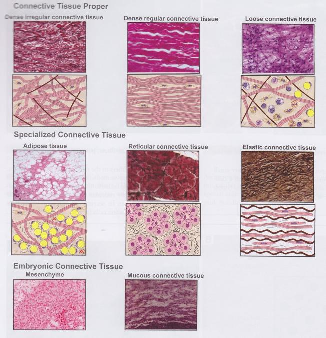
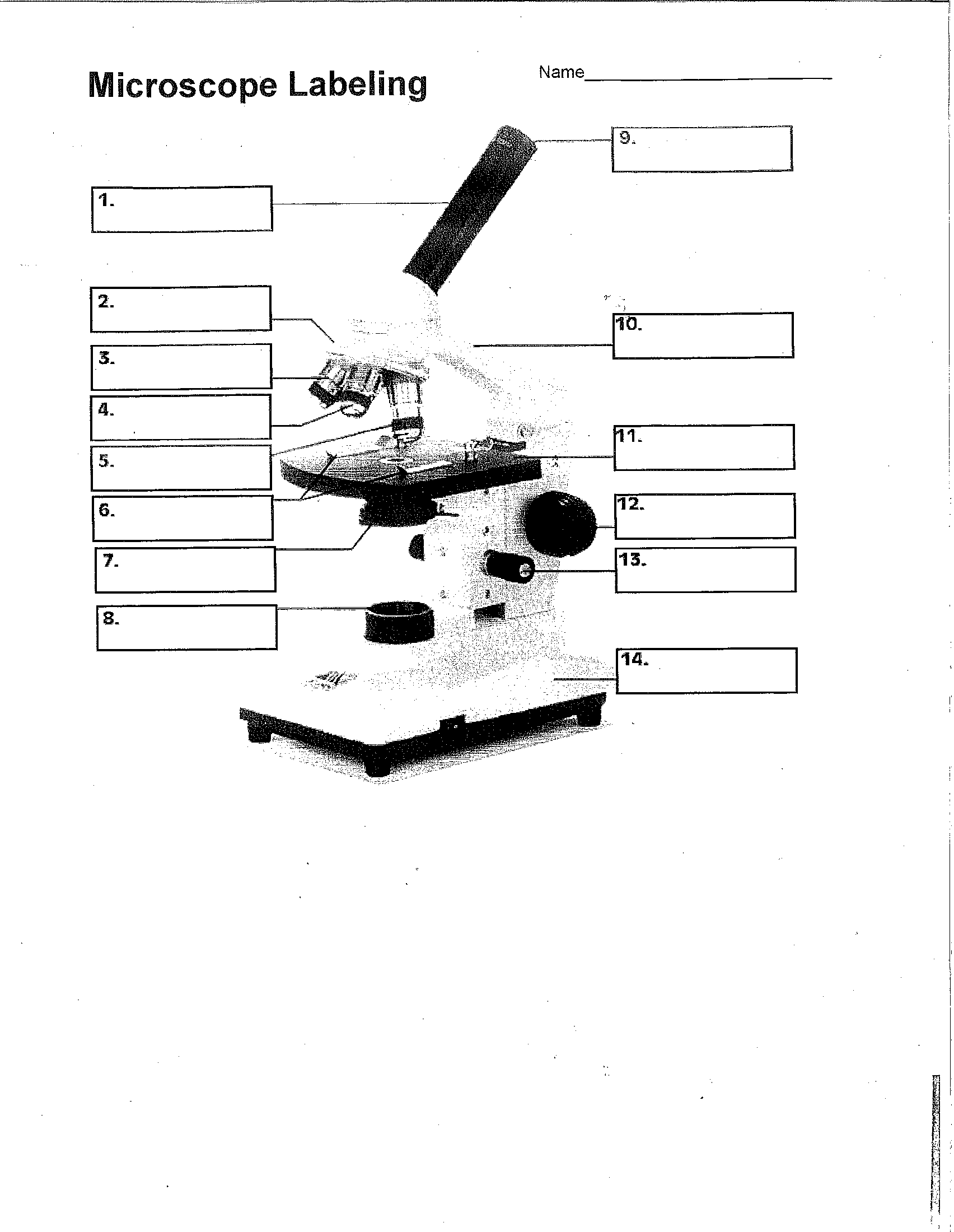
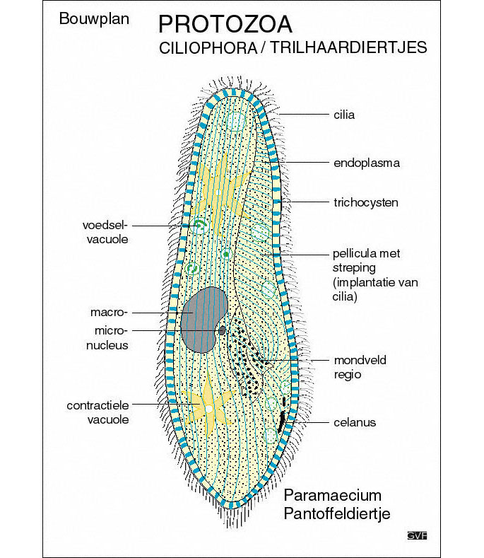
39 microscope diagram without labels
Parts of a microscope with functions and labeled diagram - Microbe Notes Figure: Diagram of parts of a microscope There are three structural parts of the microscope i.e. head, base, and arm. Head - This is also known as the body. It carries the optical parts in the upper part of the microscope. Base - It acts as microscopes support. It also carries microscopic illuminators. Free Microscope Worksheets for Simple Science Fun for Your Students 1. Parts of a Microscope . The first worksheet labels the different parts of a microscope, including the base, slide holder, and condenser. If you have a microscope, compare and contrast this worksheet to it.Also, your kids can color this microscope diagram in and read the words to each part of the microscope.
16 Parts of a Compound Microscope: Diagrams and Video Once you have an understanding of the parts of the microscope it will be much easier to navigate around and begin observing your specimen, which is the fun part! The 16 core parts of a compound microscope are: Head (Body) Arm Base Eyepiece Eyepiece tube Objective lenses Revolving Nosepiece (Turret) Rack stop Coarse adjustment knobs

Microscope diagram without labels
7th grade Science - Microscope Diagram | Quizlet The Parts of a Microscope. 12 terms. totobear PLUS. Sets found in the same folder. Science Key terms 7th grade. 13 terms. palocastillo. 7th Grade Earth Science. 9 terms. EliseC17. 7thGrade Review - Cells/Biology. 26 terms. SolizScience TEACHER. 7th grade Science, Cell theory. 8 terms. Super1412. Other sets by this creator. brain diagram without labels brain diagram without labels. Eye with labels clip art at clker.com. Neuron worksheet synapse label diagram blank worksheeto cell nerve via motor brain. Microscope with labels clip art at clker.com. Eye With Labels Clip Art at Clker.com - vector clip art online, royalty. 11 Pics about Eye With Labels Clip Art at Clker.com - vector clip art ... PDF Label parts of the Microscope: Answers Label parts of the Microscope: Answers Coarse Focus Fine Focus Eyepiece Arm Rack Stop Stage Clip . Created Date: 20150715115425Z ...
Microscope diagram without labels. Compound Microscope Parts - Labeled Diagram and their Functions The eyepiece (or ocular lens) is the lens part at the top of a microscope that the viewer looks through. The standard eyepiece has a magnification of 10x. You may exchange with an optional eyepiece ranging from 5x - 30x. [In this figure] The structure inside an eyepiece. The current design of the eyepiece is no longer a single convex lens. Label A Microscope Teaching Resources | Teachers Pay Teachers The 13 parts of the microscope: microscope, base, arm, inclination joint, course adjustment, fine adjustment, body tube, ocular lens, revolving nose piece, objectives, stage, stage clips, and iris diaphragm.Includes:13 cards with labels13 cards without labels13 labels1 blackline masterCards with labels are approx. 3¾" x 4", cards without ... blank cell diagram to label microscope diagram unlabeled light parts blank biology science labeled lab sketch worksheet labels worksheets timvandevall drawing quiz chemistry microscopes fill Mitosis Diagram Without Labels For Kids - Simple Animal Cell Unlabelled peroxisome Cell Diagram To Label - Pensandpieces pensandpieces.blogspot.com ixl organelle Compound Microscope Parts, Functions, and Labeled Diagram The individual parts of a compound microscope can vary heavily depending on the configuration & applications that the scope is being used for. Common compound microscope parts include: Eyepiece (ocular lens) with or without Pointer: The part that is looked through at the top of the compound microscope. Eyepieces typically have a magnification ...
Compound Microscope: Definition, Diagram, Parts, Uses, Working ... - BYJUS A compound microscope is defined as. A microscope with a high resolution and uses two sets of lenses providing a 2-dimensional image of the sample. The term compound refers to the usage of more than one lens in the microscope. Also, the compound microscope is one of the types of optical microscopes. The other type of optical microscope is a ... Label the Microscope Diagram | Download Scientific Diagram - ResearchGate Download scientific diagram | Label the Microscope Diagram from publication: Laboratory Exercises in Microbiology: Discovering the Unseen World through Hands-on Investigation | Microbiology ... Simple Microscope - Parts, Functions, Diagram and Labelling A simple microscope is a device that only has one lens for magnification. It functions the same way as the magnifying glass. Although it is simple in terms of design and function, it is useful I various fields including medicine, jewelry and watchmaking, and agriculture, to name a few. References A Study of the Microscope and its Functions With a Labeled Diagram ... Here, unlabeled microscope diagrams have been provided for your perusal, which will help you practice and test your understanding of the instrument. Types of Microscopes Depending on the source of illumination, microscopes can be divided into two categories. They are:
Label the microscope — Science Learning Hub In this interactive, you can label the different parts of a microscope. Use this with the Microscope parts activity to help students identify and label the main parts of a microscope and then describe their functions. Drag and drop the text labels onto the microscope diagram. Label Microscope Diagram - EnchantedLearning.com Using the terms listed below, label the microscope diagram. arm - this attaches the eyepiece and body tube to the base. base - this supports the microscope. body tube - the tube that supports the eyepiece. coarse focus adjustment - a knob that makes large adjustments to the focus. diaphragm - an adjustable opening under the stage, allowing ... PDF Parts of a Microscope Printables - Homeschool Creations Label the parts of the microscope. You can use the word bank below to fill in the blanks or cut and paste the words at the bottom. ... without needing to move the microscope ? the head •What is the magnification level on the eyepiece of a microscope?10x (see objective Microscope Parts and Functions First, the purpose of a microscope is to magnify a small object or to magnify the fine details of a larger object in order to examine minute specimens that cannot be seen by the naked eye. Here are the important compound microscope parts... Eyepiece: The lens the viewer looks through to see the specimen.
Microscope Labeled Diagram - Micropedia
How does a Microscope work A compound microscope has two or more lenses. The eyepiece or ocular lens sits atop the body tube. Many microscopes are binocular and have two ocular lenses. Additionally, a binocular head will have a prism, either in the head or the body tube, to split the image and direct it to both oculars.

All Saints Online: Diagram for Labelling: Microscope
Microscope Label Interactive Worksheets & Teaching Resources | TpT Microscope Interactive Notebook Activity by Jodi's Jewels 12 $1.89 PDF Students will complete a timeline of the history of the microscope, label a diagram, and create a pocket foldable with terms and definition cards. The timeline can be completed according to the teacher's directions or like the answer key example.

Microscope With Labels Clip Art at Clker.com - vector clip art online, royalty free & public domain
Amazing 27 Things Under The Microscope With Diagrams - Microbe Notes Under a high-power microscope, the cell organelles are more differentiated and allow the observation of individual structures. Because of the affinity of the stain with the DNA and RNA of the cell, the components inside the nucleus might also be visible. 10. DNA under the microscope.
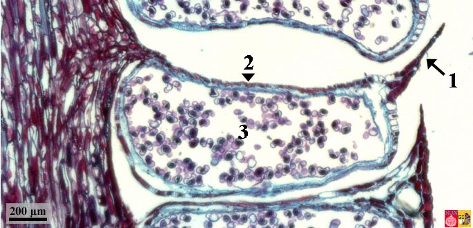
Vertebrate Histology Exam 2 Flashcards | Easy Notecards
Microscope Diagram - cell division of e coli with continuous media flow ... Microscope Diagram - 15 images - give a well labelled diagram of compound microscope using of typical, bio tem biological transmission electron microscope university, labelled microscope diagram gcse micropedia, a compound microscope diagram micropedia,
Unit 2: Cell Biology - Mrs. Frump's Classes
Microscope Labeling - The Biology Corner Students label the parts of the microscope in this photo of a basic laboratory light microscope. Can be used for practice or as a quiz. ... The type of microscope used in most science classes is the _____ microscope. 18. You should carry the microscope by the _____ and the _____. 19. The objectives are attached to what part of the microscope ...
Search in gallery
Diagram of a Compound Microscope - Biology Discussion 1. It is noted first that which objective lens is in use on the microscope. 2. Stage micrometer is positioned in such a way that it is in the field of view. 3. The eyepiece is rotated so that the two scales, the eyepiece or ocular scale and the stage micrometer scale, are parallel. 4.

Quia - 9AP Chapter 12 - The Cell Cycle (Detailed)
Labeling the Parts of the Microscope | Microscope World Resources Labeling the Parts of the Microscope This activity has been designed for use in homes and schools. Each microscope layout (both blank and the version with answers) are available as PDF downloads. You can view a more in-depth review of each part of the microscope here. Download the Label the Parts of the Microscope PDF printable version here.

basic animal cell diagram with labels
Labelled Diagram of Compound Microscope - biologydiscussion.com The below mentioned article provides a labelled diagram of compound microscope. Part # 1. The Stand: The stand is made up of a heavy foot which carries a curved inclinable limb or arm bearing the body tube. The foot is generally horse shoe-shaped structure (Fig. 2) which rests on table top or any other surface on which the microscope in kept.

microscope labeled microscope worksheet labeling sc 1 st template entrancing labelling - Top ...
Parts of Stereo Microscope (Dissecting microscope) - labeled diagram ... Stereo microscopes (also called Dissecting microscope) are branched out from other light microscopes for the application of viewing "3D" objects. These include substantial specimens, such as insects, feathers, leaves, rocks, sand grains, gems, coins, and stamps, etc. Functionally, a stereo microscope is like a powerful magnifying glass.

Microscope - Science
Microscope, Microscope Parts, Labeled Diagram, and Functions The Iris Diaphragm is located above the condenser lens and below the microscope stage. The different sized holes in the diaphragm helps to vary the size of the cone and intensity of light that is projected upward into the slide. However, there is no set rule regarding which setting to use for a particular power.

Microscope labeled diagram
Parts of the Microscope with Labeling (also Free Printouts) A microscope is one of the invaluable tools in the laboratory setting. It is used to observe things that cannot be seen by the naked eye. Table of Contents 1. Eyepiece 2. Body tube/Head 3. Turret/Nose piece 4. Objective lenses 5. Knobs (fine and coarse) 6. Stage and stage clips 7. Aperture 9. Condenser 10. Condenser focus knob 11. Iris diaphragm
Search in gallery
PDF Label parts of the Microscope: Answers Label parts of the Microscope: Answers Coarse Focus Fine Focus Eyepiece Arm Rack Stop Stage Clip . Created Date: 20150715115425Z ...
Bean | Free Images at Clker.com - vector clip art online, royalty free & public domain
brain diagram without labels brain diagram without labels. Eye with labels clip art at clker.com. Neuron worksheet synapse label diagram blank worksheeto cell nerve via motor brain. Microscope with labels clip art at clker.com. Eye With Labels Clip Art at Clker.com - vector clip art online, royalty. 11 Pics about Eye With Labels Clip Art at Clker.com - vector clip art ...

Easy Microscope Diagram With Labels - Micropedia
7th grade Science - Microscope Diagram | Quizlet The Parts of a Microscope. 12 terms. totobear PLUS. Sets found in the same folder. Science Key terms 7th grade. 13 terms. palocastillo. 7th Grade Earth Science. 9 terms. EliseC17. 7thGrade Review - Cells/Biology. 26 terms. SolizScience TEACHER. 7th grade Science, Cell theory. 8 terms. Super1412. Other sets by this creator.

Label the Microscope Part

Microscope - 7 Red Team
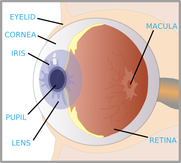
Eye With Labels Clip Art at Clker.com - vector clip art online, royalty free & public domain
Post a Comment for "39 microscope diagram without labels"