44 basic animal cell diagram with labels
Venn diagram - Wikipedia A Venn diagram is a widely used diagram style that shows the logical relation between sets, popularized by John Venn (1834–1923) in the 1880s. The diagrams are used to teach elementary set theory, and to illustrate simple set relationships in probability, logic, statistics, linguistics and computer science.A Venn diagram uses simple closed curves drawn on a plane to represent sets. Printable Animal Cell Diagram - Labeled, Unlabeled, and Blank Printable animal cell diagram to help you learn the organelles in an animal cell in preparation for your test or quiz. 5th grade science and biology. Soul Candy Inter. Design (253) 376-9675 Homeschool
IXL | Animal cell diagrams: label parts | 5th grade science Improve your science knowledge with free questions in "Animal cell diagrams: label parts" and thousands of other science skills.

Basic animal cell diagram with labels
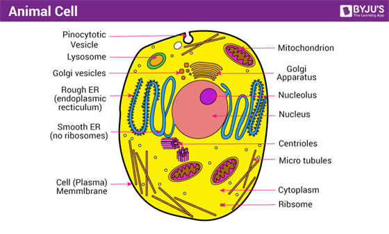
Animal Cell - Structure, Function, Diagram and Types - BYJUS The most common types of animal cells are: Skin Cells Melanocytes, keratinocytes, Merkel cells and Langerhans cells Muscle Cells Myocyte, Myosatellite cells, Tendon cells, Cardiac muscle cells Blood Cells Leukocytes, erythrocytes, platelet Nerve Cells Schwann cell, glial cells etc Fat Cells Adipocytes Points to Note About Animal Cell Cell Worksheets | Plant and Animal Cells - Math Worksheets 4 Kids This collection of animal and plant cell worksheets strikes a balance between cognitive and psychomotor domains of learning and offers a conceptual grounding in cell biology. The worksheets recommended for students of grade 4 through grade 8 feature labeled animal and plant cell structure charts and cross-section charts, cell vocabulary with ... The Mitochondrion - Molecular Biology of the Cell - NCBI Bookshelf The cell was stained with a fluorescent dye (rhodamine 123) that specifically labels mitochondria Figure 14-6 ... In this diagram, the high-energy electrons are shown as two red dots on a yellow hydrogen atom. A hydride ion (H-a hydrogen atom and an extra electron) is removed from NADH and is converted into a proton and two high-energy . Figure 14-10. A summary of energy …
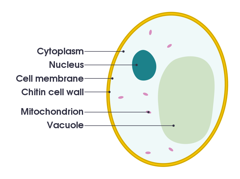
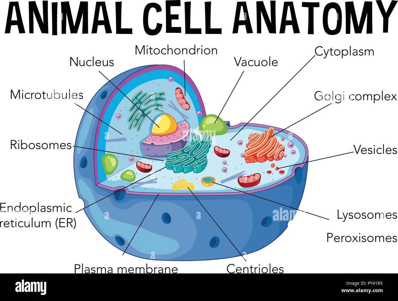
Basic animal cell diagram with labels. Animal Cell Diagram | Science Trends An animal cell diagram is a great way to learn and understand the many functions of an animal cell. The diagram, like the one above, will include labels of the major parts of an animal cell including the cell membrane, nucleus, ribosomes, mitochondria, vesicles, and cytosol. A Well-labelled Diagram Of Animal Cell With Explanation - BYJUS Well-Labelled Diagram of Animal Cell The Cell Organelles are membrane-bound, present within the cells. There are various organelles present within the cell and are classified into three categories based on the presence or absence of membrane. Listed below are the Cell Organelles of an animal cell along with their functions. Animal Cells: Labelled Diagram, Definitions, and Structure - Research Tweet Animal Cells Organelles and Functions. A double layer that supports and protects the cell. Allows materials in and out. The control center of the cell. Nucleus contains majority of cell's the DNA. Popularly known as the "Powerhouse". Breaks down food to produce energy in the form of ATP. animal cells without labels Animal Cell Labelling Activity | Basic Animal Cell Diagram. 15 Pictures about Animal Cell Labelling Activity | Basic Animal Cell Diagram : 30 Label Of Animal Cell, Animal cell - 3D - YouTube and also visual aid project for cells - Google Search | Science/Physics/Energy.
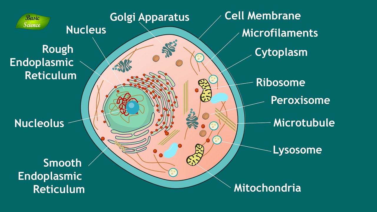
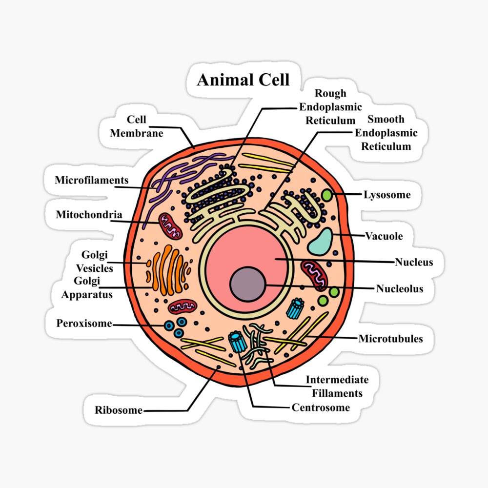
Label the Animal Cell - EnchantedLearning.com Golgi body - (also called the Golgi apparatus or Golgi complex) a flattened, layered, sac-like organelle that looks like a stack of pancakes and is located near the nucleus. It produces the membranes that surround the lysosomes. The Golgi body packages proteins and carbohydrates into membrane-bound vesicles for "export" from the cell. Animal Cell Basic Diagram : Animal Cell Structure Function Diagram And ... Molecular Expressions Cell Biology Animal Cell Structure from micro.magnet.fsu.edu The plasma membrane structure is made up of several components, depending on the type of cell you're looking at, but they all share one major component: Labeled diagram of plant cell, created with biorender.com. May 23, 2021 · animal cell culture requires the ... Guidelines for Safe Work Practices in Human and Animal Medical ... 06.01.2012 · Regardless, animal cadavers can harbor zoonotic agents, and risk assessment to determine whether zoonotic infectious agents may be present in a cadaver, as outlined in Section 12, is critically important for establishing appropriate animal necropsy biosafety procedures. The guidelines in this section are combined biosafety best practices for both human autopsy and … Free Printable Plant and Animal Cells Worksheets - Homeschool Giveaways Label the Parts of an Animal Cell. Printable Label and Color the Parts Animal Cell - This animal cell handout is perfect for students studying the different parts of the cell including the SER (Smooth Endoplasmic Reticulum), Golgi Bodies, and more.. Label the Animal Cell Worksheets - Grab these set of cell diagrams for students to label themselves and as topics of discussion during your ...
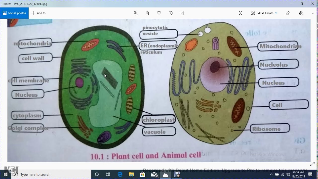
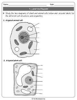
Animal Cell: Animal Cell Diagram: Types, and Functions - Embibe An animal cell is a eukaryotic cell having membrane-bound cell organelles without a cell wall. The cell size varies from a few microns to a few centimetres. For example, the largest animal cell is the ostrich egg measuring 170 mm x 130 mm. We can say that the size of the cell depends on the function it performs. Plant Cells Vs. Animal Cells (With Diagrams) - Owlcation The most important structures of plant and animal cells are shown in the diagrams below, which provide a clear illustration of how much these cells have in common. The significant differences between plant and animal cells are also shown, and the diagrams are followed by more in-depth information. Diagram of an animal cell Doc Sonic Animal Cell/Plant Cell Structure Diagram Printable (Blank) - TeacherVision Ready-to-label cell diagrams for tests, homework, quizzes, and study aids. This printable is the perfect way to test students' knowledge of cellular biology. Featuring blank diagrams of an animal cell and a plant cell, plus plenty of space for labels and notes, it's perfect for use as a study aid, quick quiz, homework assignment, or biology test. Animal cells - Cell structure - AQA - BBC Bitesize Animal cells have a basic structure. Below the basic structure is shown in the same animal cell, on the left viewed with the light microscope, and on the right with the transmission electron...
ArtStation - KS3 Animal Cell Worksheet
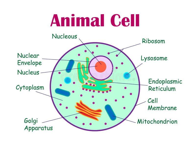
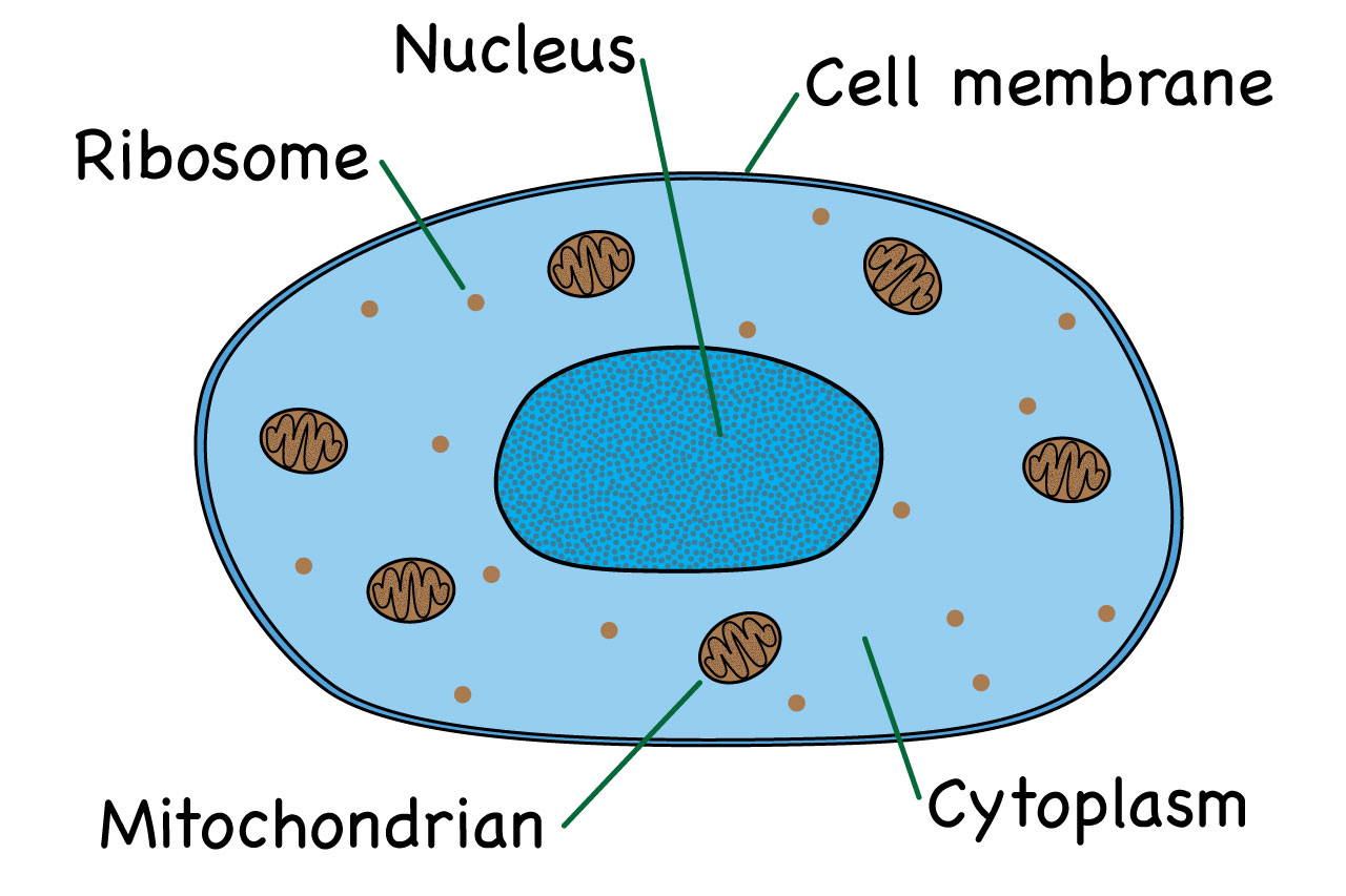
Animal Cell- Definition, Structure, Parts, Functions, Labeled Diagram Definition of animal cell An animal cell is a eukaryotic cell that lacks a cell wall, and it is enclosed by the plasma membrane. The cell organelles are enclosed by the plasma membrane including the cell nucleus. Unlike the animal cell lacking the cell wall, plant cells have a cell wall.
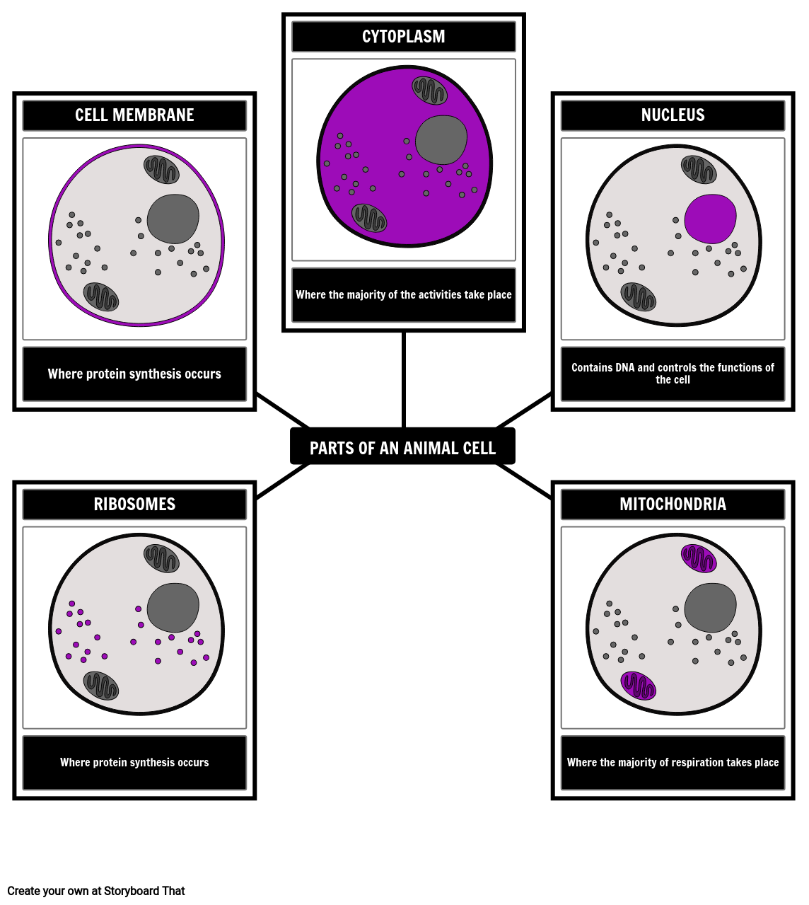
Parts of an Animal Cell | Label an Animal Cell Activity
(PDF) Basic Virology, Third Edition - Academia.edu Basic Virology, Third Edition. Abdu Abdoulaye. Download Download PDF. Full PDF Package Download Full PDF Package. This Paper. A short summary of this paper. 37 Full PDFs related to this paper. Download. PDF Pack. People also downloaded these PDFs. People also downloaded these free PDFs. People also downloaded these free PDFs . Edward K. Wagner, Martinez J. …
Animal Cell Stock Illustrations – 11,296 Animal Cell Stock ...
Interactive Bacteria Cell Model - CELLS alive Periplasmic Space: This cellular compartment is found only in those bacteria that have both an outer membrane and plasma membrane (e.g. Gram negative bacteria).In the space are enzymes and other proteins that help digest and move nutrients into the cell. Cell Wall: Composed of peptidoglycan (polysaccharides + protein), the cell wall maintains the overall shape of a …

Animal Cell- Definition, Structure, Parts, Functions, Labeled ...
Animal and Plant Cell Worksheets - Super Teacher Worksheets Plant Cell Parts (Color Poster) FREE This is a basic illustration of a plant cell with major parts labeled. Labels include nucleus, chloroplast, cytoplasm, membrane, cell wall, and vacuole, and mitochondrion. Use it as a poster in your classroom or have students glue it into their science notebooks. View PDF Plant Cell Vocabulary Cards
25 Major Difference Between Plant Cell and Animal Cell
Animal Cell diagram with labels by Russell Kightley Media Labelled Animal Cell Diagram. ( plant cells are somewhat different). A cell is a complete functional biological unit with many different internal structures. Somewhat like an entire city in miniature. It is enclosed by a cell membrane and has a nucleus (shown in purple) in the middle. Cells are the building blocks of our bodies.
Draw a neat labelled diagram of an animal cell (with ...
Animal Cell Parts - Biology Wise The labeled diagram given below depicts the parts of an animal cell, which will help you in understanding the concept better. Labeled Animal Cell Diagram What Are the Various Parts of an Animal Cell? Cell Membrane: The cell membrane is the outermost part of the cell, which encloses all the other cell organelles.
What Is An Animal Cell? Facts, Pictures & Info For Kids ...
A Labeled Diagram of the Animal Cell and its Organelles One can observe the golgi apparatus in the labeled animal cell parts diagram. The golgi apparatus is situated near the cell nucleus and besides the stacked sacs, it also contains large number of vesicles. The main function of this golgi complex is to receive the proteins synthesized in the ER and transform it into more complex proteins.

1.4 Summary | Cells as the basic units of life | Siyavula
Featured picture candidates/Cell membrane (diagrammatic) Edit 3 uploaded Standard zoom boxes tend to obscure the labels, but if there is a consensus to change to those, I'll give it another try. Dhatfield ( talk ) 20:29, 16 June 2008 (UTC) Reply [ reply ] Strong oppose There are only unsaturated tails on the phospholipids, for a reduced structure diagram of a cell membrane's lipid bilayer there should be one unsaturated and one saturated …
A Well-labelled Diagram Of Animal Cell With Explanation
The Cell - ScienceQuiz.net Look at the diagram of an animal cell. Select correct statement from the following about animal cells. Select correct statement from the following about animal cells. A is the cell membrane and DNA is located inside B.

Basics of Animal Cell Biology | LoveToKnow
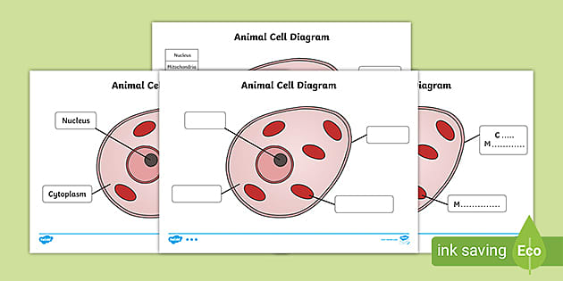
Label the Animal Cell: Level 1 | Worksheet | Education.com In Label the Animal Cell: Level 1, students will use a word bank to label the parts of a cell in an animal cell diagram. To take the learning one step further, have students assign a color to each of the organelles and then color in the diagram. For a broader focus, use this worksheet in conjunction with the Label the Plant Cell: Level 1 worksheet.

A Labeled Diagram of the Animal Cell and its Organelles ...
An Overview of Hyphae in Fungi, Their Function and Types. 10.01.2022 · Fungi have their cell wall made up of chitin. Their body is composed of long thread-like filaments or tubes known as hyphae . In singular form, this structure is referred to as a hypha.
How to draw a typical animal cell - Quora
Label Cell Parts | Plant & Animal Cell Activity | StoryboardThat Create a cell diagram with each part of plant and animal cells labeled. Include descriptions of what each organelle does. Click "Start Assignment". Find diagrams of a plant and an animal cell in the Science tab. Using arrows and Textables, label each part of the cell and describe its function.

File:Differences between simple animal and plant cells (en ...
Animal and plant cell labeling - Teaching resources - Wordwall Animal and plant cell card sort Group sort. by Braesbiology. Plant and Animal Cell GCSE Recap Labelled diagram. by Seraroberts. Basic Plant and Animal Cell Structure Labelled diagram. by Kate92. Label a plant and animal cell Labelled diagram. by Jenniferross. Y10 Biology.
plant cell and animal cell diagram for class 8 | plant cell and animal cell labeled diagram
Junqueira's Basic Histology Text and Atlas, 14th Edition Junqueiras Basic Histology Text and Atlas 14th Edition Vet Books ir. by Zelle Peredas. Download Free PDF Download PDF Download Free PDF View PDF. Basic Histology. by MARIAN ESTRADAR. Download Free PDF Download PDF Download Free PDF View PDF. Color Atlas of Cytology, Histology, and Microscopic Anatomy. by Mina Ungureanu. Download Free PDF Download PDF …

Animal Cell Diagram | Woo! Jr. Kids Activities : Children's ...
Free Animal Cell Diagram Templates - Edrawsoft A clear design animal cell diagram template from Edraw is waiting for you in the free download version. Use it for any kinds of science coursework or group discussions. You can also adjust the diagram sizes at any time you want for more insights.
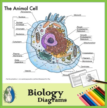
Animal Cell Diagrams for Coloring and Labeling, with Reference Chart and Summary
What Is An Animal Cell? Facts, Pictures & Info For Kids & Students. Most animal cells are between 10 and 20 micrometers across. A micrometer is one millionth of a meter, or one thousandth of a millimeter. In other words, most animal cells are very small! Although most animal cells are far too small to be seen without a microscope, some are much larger. The human egg cell, for example, is visible to the naked eye.

Plant Cells Vs. Animal Cells (With Diagrams) - Owlcation
dock8 deficiency attenuates microglia colonization in early 17.08.2022 · Microglia are tissue-resident macrophages that carry out immune functions in the brain. The deficiency or dysfunction of microglia has been implicated in many neurodegenerative disorders. DOCK8, a ...
1,117 Plant Cell Diagram Stock Photos, Pictures & Royalty ...
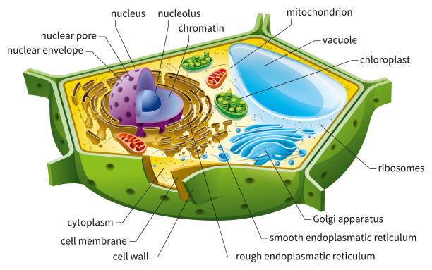
Plant and Animal Cell: Labeled Diagram, Structure, Function - Embibe Plant and Animal Cell: The cell is the basic building block of life. Cells are responsible for all aspects of life. The number of cells in an organism determines its classification. Unicellular species have only one cell, but multicellular organisms contain many cells. ... Diagram of Plant and Animal Cell. Fig: Plant Cell. Fig: Animal Cell ...
File:Simple diagram of yeast cell (en).svg - Wikipedia
Animal Cell - The Definitive Guide | Biology Dictionary Animal cells are the basic unit of life in organisms of the kingdom Animalia. They are eukaryotic cells with a defined nucleus and membrane-bound organelles ... Additionally, some organelles will be highly abundant in certain cells and not others. Labeled diagram of a typical animal cell Nucleus. The nucleus contains all the genetic material in ...
Animal Cell Structure and Function | Notes | Eukaryotic Cell | Basic Science Series
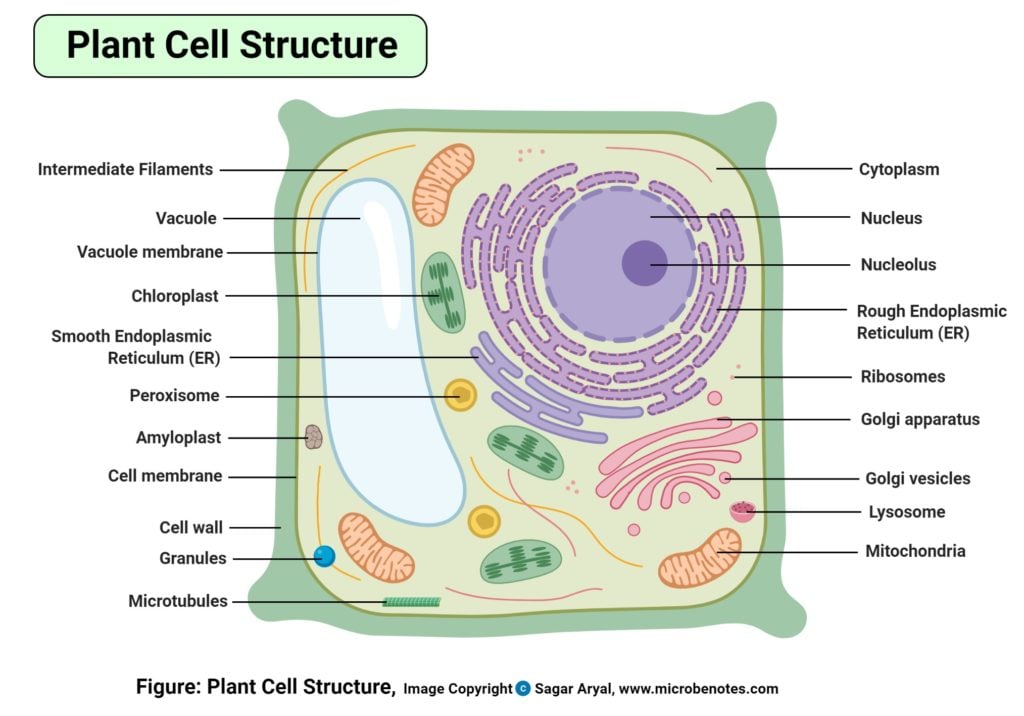
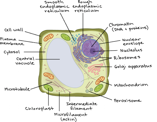
Structure of Animal Cell and Plant Cell Under Microscope Generalized Structure of a Plant Cell Diagram. As you can see in the above labeled plant cell diagram under light microscope, there are 13 parts namely, Cell membrane. Cytoplasm. Ribosomes. Nucleus. Smooth Endoplasmic Reticulum. Lysosome.
Labeled Animal Cell Diagram" Poster for Sale by BundaBear ...
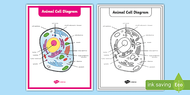
Animal Cell Labelling Activity | Basic Animal Cell Diagram - Twinkl This basic animal cell diagram with labels is simple yet provides your children with a visual aid and allows them to apply their knowledge of animals cells. With that in mind, we have created a range of resources to help those who teach their students about cells.
Animal cell label - Teaching resources
The Mitochondrion - Molecular Biology of the Cell - NCBI Bookshelf The cell was stained with a fluorescent dye (rhodamine 123) that specifically labels mitochondria Figure 14-6 ... In this diagram, the high-energy electrons are shown as two red dots on a yellow hydrogen atom. A hydride ion (H-a hydrogen atom and an extra electron) is removed from NADH and is converted into a proton and two high-energy . Figure 14-10. A summary of energy …

Animal Cell Anatomy Diagram Structure with all parts nucleus ...
Cell Worksheets | Plant and Animal Cells - Math Worksheets 4 Kids This collection of animal and plant cell worksheets strikes a balance between cognitive and psychomotor domains of learning and offers a conceptual grounding in cell biology. The worksheets recommended for students of grade 4 through grade 8 feature labeled animal and plant cell structure charts and cross-section charts, cell vocabulary with ...
Animal Cell Png - Simple Animal Cell Diagram Without Labels ...
Animal Cell - Structure, Function, Diagram and Types - BYJUS The most common types of animal cells are: Skin Cells Melanocytes, keratinocytes, Merkel cells and Langerhans cells Muscle Cells Myocyte, Myosatellite cells, Tendon cells, Cardiac muscle cells Blood Cells Leukocytes, erythrocytes, platelet Nerve Cells Schwann cell, glial cells etc Fat Cells Adipocytes Points to Note About Animal Cell

Label the Animal Cell Worksheets (SB11866) | Animal cells ...

Animal Cell Structure and Organelles with their functions ...
Animal cell diagram hi-res stock photography and images - Alamy

Animal Cell - The Definitive Guide | Biology Dictionary

Animal Cell Diagram - Tim's Printables
Animal And Plant Cell Diagram To Label Teaching Resources | TpT
Animal Cell Diagram
Animal Cell Labels Diagram | Quizlet
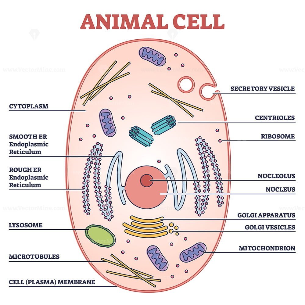
Animal cell with labeled anatomic structure parts diagram outline diagram

7,139 Plant Cell Illustrations & Clip Art - iStock

Animal Cell Diagram | Science Trends
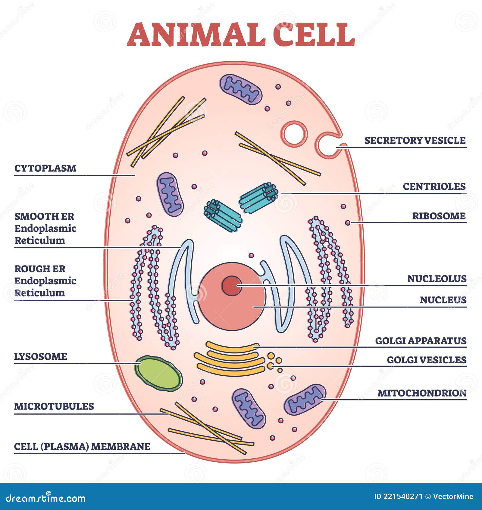
Animal Cell with Labeled Anatomic Structure Parts Diagram ...

Plant Cell - The Definitive Guide | Biology Dictionary
Animal Cell Labelling Activity | Primary Resources | Twinkl
Plant vs animal cells review (article) | Khan Academy
Printable Animal Cell Diagram | Life Science Resources | 3-5

File:Simple diagram of animal cell (en).svg - Wikimedia Commons
Simple Diagram Of Animal Cell - Simple Animal Cell Unlabelled ...

Animal Cell Structure and Organelles with their functions
What is the correct diagram of plant and animal cell? - Quora
Post a Comment for "44 basic animal cell diagram with labels"